Boston surgeons have achieved a surprising medical feat by operating on the brain of a fetus that was still in its mother’s womb. Two months after her birth, the little girl is in good health.
It is a unique surgical operation in the world which took place a few weeks ago. On March 15, surgeons in Boston (United States) managed to treat a cerebral vascular malformation – also known as an aneurysm of the vein of Galen – on a fetus which was in the womb of its mother.
As explained by our colleagues from CNN, the results of this unprecedented operation were communicated last Thursday, May 4, in the columns of the specialized journal Stroke. The operation was urgent to say the least: an aneurysm of the vein of Galen is likely to cause a “significant increase in the flow and pressure in the veins of the malformation and the brain”, explains on its website the Center Hospitalier University of Lausanne (Switzerland).
A “brain malformation” that was getting worse
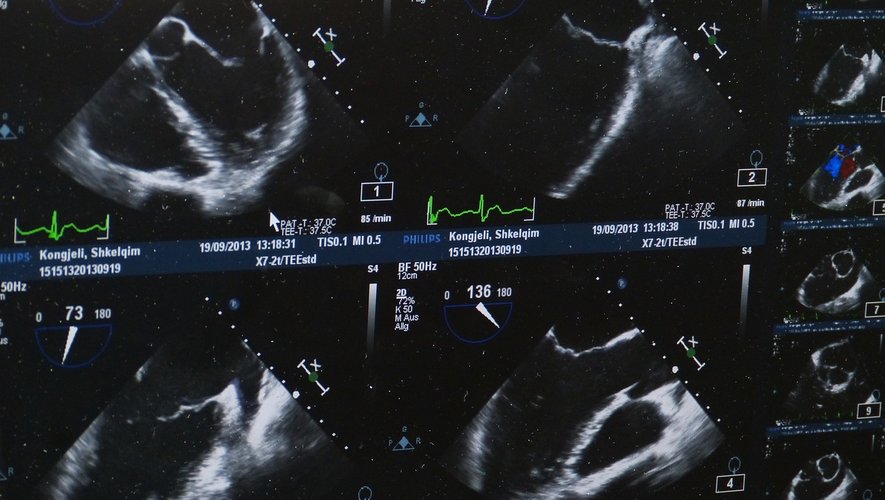
The mother was in her 30th week of pregnancy when she and her partner discovered their child’s malformation during an ultrasound session. The Brigham and Women’s Hospital and the Boston Children’s Hospital then want the young couple to take part in a clinical trial: “The baby did not yet have a heart failure problem, nor was there any brain damage. But the malformation was getting bigger and bigger,” said Carol Benson, a radiologist at Brigham and Women’s Hospital. If the doctors had decided to wait for the birth of the child to operate on it, the baby risked serious cerebral complications: “40% of the children victims of this disease die shortly after their birth”, indicates for her part the radiologist Darren Orbach, Boston Children’s Hospital, with CNN.
The operation itself required complex logistics. The fetus had to be positioned such that its head was positioned towards its mother’s abdominal wall. Doctors performed a first injection, with the aim of immobilizing the fetus. A second injection was performed to relieve the pain. Surgeons later fitted a catheter to the baby to slow blood flow and reduce blood pressure. The latter were finally guided by ultrasound throughout the operation.
The baby girl was finally born two days later. Two months later, her state of health is very satisfactory: “She is not taking any treatment, no surgery is planned. She does not need heart machines, she breathes on her own. She is eating well, she is growing “, evokes Dr. Carole Benson who describes an exceptional surgical procedure.

